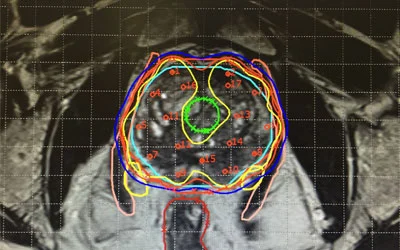
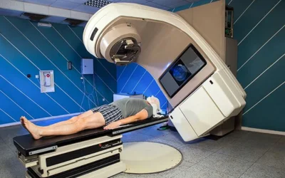
Dr. Pooja Khullar is working as Senior Consultant in Radiation Oncology at Dharamshila Narayana
Super Speciality Hospital, Delhi, and trained at Rajiv Gandhi Cancer Institute and Research
Centre, Delhi. She has 12 years of experience in superior advanced radiotherapy skills like
IMRT, IGRT, SBRT, SRT, SRS, 3D Brachytherapy, and Respiratory Gating.
MBBS – B.R.D Medical College, Gorakhpur (UP)
DNB (Radiation Oncology) – Rajiv Gandhi Cancer Institute & Research Centre, Delhi
Short term Training – Dept. of Radiotherapy, AIIMS, New Delhi
Short term Training – Sanjay Gandhi PGI, Lucknow
Fellowship – ESMO Translational Research Unit, Naples, Italy (2016)
3D Brachytherapy
3D brachytherapy is an advanced medical technique employed in the treatment of various cancers, particularly prostate cancer and gynecological malignancies. Unlike traditional brachytherapy, which relies on two-dimensional imaging, 3D brachytherapy utilizes three-dimensional imaging technology, such as CT or MRI scans, to precisely target cancerous tissues while minimizing damage to surrounding healthy tissue. This approach allows for more accurate placement of radiation sources directly into the tumor, resulting in higher treatment efficacy and reduced side effects. By customizing the radiation dose based on the tumor’s specific size, shape, and location, 3D brachytherapy offers patients a tailored and effective treatment option, often leading to improved outcomes and better quality of life.
3D brachytherapy revolutionizes cancer treatment by combining precision targeting with advanced imaging technology. Unlike traditional methods, it utilizes detailed three-dimensional models of tumors and surrounding tissues, allowing oncologists to tailor treatment plans to each patient’s unique anatomy. This approach minimizes radiation exposure to healthy tissues, reducing side effects and improving outcomes. With enhanced dose delivery and real-time monitoring, 3D brachytherapy ensures patient safety while expanding treatment options for a wide range of cancers, from prostate to gynecological. Its ability to deliver precise radiation doses directly to tumors makes it a valuable tool in the fight against cancer, offering hope and improved quality of life for patients.
Precision targeting minimizes side effects.
Customized plans tailored to each patient.
Expands treatment options for various cancers.
Enhanced dose delivery improves outcomes.
Advanced imaging ensures patient safety.
Precision targeting minimizes radiation exposure.
Respiratory Gating and Deep Inspiration Breath Hold (DIBH)
Accurately tracking tumor position is a critically important factor when maximizing radiation dose to the tumor and limiting normal tissue exposure.
What is respiratory gating?
Gating is a system that tracks a patient’s normal respiratory cycle with an infrared camera and chest/abdomen marker. The system is coordinated to only deliver radiation when the tumor is in the treatment field.
The respiratory gating radiotherapy technique can apply to moving tumors, such lung and liver cancer. Real-time Position ManagementTM(RPM) respiratory gating system, which gates the beam delivery during treatment to account for organ motion.
The infrared camera tracks tumors that move during treatment as the patient breathes in and out, then the linac accelerator controlled x -ray beam treats the tumor within the gated phase to assure the accuracy of treatment position. The non-invasive infrared tracking marker box is placed on patient’s body. We have the big bore CT simulator with four[1]dimensional CT capability, it can record combines continuous monitor chest wall movement during treatment with pre-plotted target trajectories to ensure the maximum dose is delivered to the target while avoiding critical organs. In this case, we can treat patients with high accuracy while minimize treatment errors.
Radiation beam is gated to when the target falls within the treatment field, patient can breathe normally and remain comfortable and high compliance during the treatment. This technique will takes about five to ten minutes more than the regular treatment time.
What can a patient expect when treated with the respiratory-gated system?
The patient is first imaged on the CT Simulator while tracking the movement of the lightweight chest/abdomen marker with the infrared camera. Images are analyzed using 4D software (the fourth dimension being time) to accurately determine tumor motion relating to breathing.
When the patient arrives for treatment, the chest/abdomen marker will be placed in the same location as set for imaging. The infrared camera will track the patient’s breathing cycle as he or she is allowed to breathe in a relaxed manner. The gating system will automatically detect when to give radiation according to the patient’s specific respiratory cycle. The beam will be held off for disruptions in the breathing cycle, such as coughing or sneezing. Using the respiratory gating system, the patient will receive better tumor control and less normal tissue complications without sacrificing comfort.
What is DIBH treatment?
Respiratory Deep Inspiration Breath Hold (DIBH) is a specific radiation therapy technique for Breast treatment to spare doses to the heart and lungs. Using the DIBH technique, the radiation is delivered only at certain points during the patient’s breathing cycle of inspiration and expiration. The patient is asked to take a deep breath in and hold their breath for about 20 seconds.
This, in turn, will limit the amount of the heart and lung that is exposed to the radiation beam, since taking a deep breath in will allow these organs to move out of the treatment field (See sample picture of heart position completely out of the yellow line with DIBH). DIBH can be also used to minimize internal organ motion for other body sites, such as the stomach, pancreas, and liver.
Stereotactic Radiation Therapy (SRT) and Stereotactic Radiosurgery (SRS) represent groundbreaking advancements in the field of oncology, offering precise and targeted treatment for various types of cancerous and non-cancerous conditions. SRT and SRS utilize highly focused beams of radiation to precisely deliver therapeutic doses to tumors while minimizing damage to surrounding healthy tissues.
SRT involves the administration of radiation therapy in multiple fractions, typically over several days or weeks. It’s particularly effective for treating larger tumors or those located near critical structures where minimizing radiation exposure is crucial. This fractionated approach allows for the delivery of higher total doses while reducing the risk of toxicity to healthy tissues.
On the other hand, SRS is a non-invasive procedure that delivers a single, high dose of radiation to a specific target in a single session. Unlike traditional surgery, which involves physically removing the tumor, SRS achieves tumor destruction through precise targeting of radiation beams. It’s commonly used for small to medium-sized tumors, especially those located in the brain, spine, and certain other areas where surgical intervention may be challenging.
Both SRT and SRS rely on advanced imaging techniques, such as MRI, CT scans, and/or PET scans, to precisely localize the tumor and guide the delivery of radiation with submillimeter accuracy. This precise targeting minimizes radiation exposure to surrounding healthy tissues, reducing the risk of side effects and improving treatment outcomes.
These treatment modalities offer several advantages over traditional radiation therapy and surgery. Firstly, they are less invasive, which means shorter recovery times and reduced risk of complications. Additionally, they can be used to treat tumors that may be considered inoperable or too risky for conventional surgery. Furthermore, SRT and SRS are highly effective in controlling or eliminating tumors, leading to improved long-term survival rates and quality of life for patients.
SRT and SRS have revolutionized the management of various cancers and other medical conditions, including brain tumors, spinal tumors, lung cancer, liver cancer, prostate cancer, and metastatic disease. They are often used as primary treatments, adjuvant therapies, or palliative measures, depending on the specific circumstances of each case.
Stereotactic Body Radiation Therapy (SBRT) represents a cutting-edge approach in the field of radiation oncology, offering precise and potent treatment for various cancers with minimal damage to surrounding healthy tissue. This innovative technique delivers high doses of radiation to tumors with pinpoint accuracy, typically in just a few sessions, making it an attractive option for patients seeking effective treatment with fewer side effects and shorter treatment times.
At the heart of SBRT lies advanced imaging technology, such as CT scans, MRI, or PET scans, which allows oncologists to precisely visualize the tumor and surrounding structures in three dimensions. This detailed imaging is crucial for accurately targeting the tumor and minimizing radiation exposure to nearby organs and tissues.
One of the key advantages of SBRT is its ability to complete treatment in just a few sessions, often as few as one to five sessions, compared to conventional radiation therapy, which may require daily treatments over several weeks. This condensed treatment schedule not only reduces the burden on patients but also minimizes disruptions to their daily lives, allowing them to resume normal activities sooner.
Furthermore, SBRT is particularly well-suited for treating tumors in challenging locations, such as those near critical structures like the spinal cord, liver, or lungs. Its precise targeting capabilities enable oncologists to deliver high doses of radiation to these tumors while minimizing the risk of damage to nearby healthy tissues, thereby expanding the range of cancers that can be effectively treated with radiation therapy.
The effectiveness of SBRT has been demonstrated across various cancer types, including lung cancer, liver cancer, prostate cancer, and pancreatic cancer, among others. Numerous studies have shown high rates of local tumor control and excellent long-term outcomes for patients treated with SBRT, making it a valuable addition to the oncologist’s arsenal of treatment options.
Despite its effectiveness, SBRT may not be suitable for all patients or all types of tumors. Factors such as tumor size, location, and proximity to critical structures must be carefully considered when determining whether SBRT is the appropriate treatment approach. Additionally, like any form of radiation therapy, SBRT carries some risk of side effects, which may include fatigue, skin irritation, and, in rare cases, damage to nearby organs.
Image-guided radiation therapy (IGRT) revolutionizes cancer treatment by enhancing precision and accuracy, thereby minimizing damage to healthy tissues surrounding tumors. This advanced form of radiation therapy integrates imaging techniques with radiation delivery, allowing for real-time visualization and adjustment during treatment sessions. By precisely targeting tumors while sparing healthy tissue, IGRT significantly improves therapeutic outcomes and reduces potential side effects.
At the core of IGRT lies its ability to utilize various imaging modalities to precisely locate tumors and monitor their position throughout the treatment process. Techniques such as X-rays, CT scans, MRI, and PET scans are employed to create detailed images of the tumor and its surrounding anatomy. These images serve as a guide for radiation oncologists to accurately deliver radiation to the tumor site while minimizing exposure to nearby critical structures.
One of the key benefits of IGRT is its ability to account for anatomical changes that may occur during the course of treatment. Tumors and surrounding tissues can shift or change shape due to factors such as organ motion, patient positioning, or changes in the tumor itself. IGRT allows clinicians to adapt treatment plans in real-time based on these changes, ensuring that the radiation is consistently delivered to the intended target with high precision.
The integration of imaging technology into the radiation therapy process also enables clinicians to verify the position of the patient and the tumor immediately before each treatment session. This verification step, known as image guidance, ensures that the patient is in the correct position and that the radiation beams are accurately aligned with the tumor. Any necessary adjustments can be made on-the-spot, further enhancing the accuracy of treatment delivery.
IGRT is particularly beneficial for tumors located in areas of the body that are prone to movement or are surrounded by critical structures, such as the lungs, liver, prostate, and head and neck region. By precisely tracking the tumor’s position and adjusting for movement, IGRT allows for higher radiation doses to be delivered to the tumor while minimizing the risk of damaging nearby healthy tissues. This can result in better tumor control and improved patient outcomes.
In addition to its precision and accuracy, IGRT offers the potential for shorter treatment times and reduced toxicity compared to conventional radiation therapy techniques. By delivering higher doses of radiation more precisely, IGRT may also improve overall treatment efficacy and increase the likelihood of tumor eradication.
Intensity-modulated radiation therapy (IMRT) stands as a pioneering advancement in cancer treatment, revolutionizing how radiation is delivered with precision and effectiveness. This innovative technique tailors radiation beams to conform closely to the shape of the tumor, sparing healthy surrounding tissues from unnecessary exposure while delivering higher doses of radiation to the cancerous cells.
At the heart of IMRT lies its ability to modulate the intensity of radiation beams across multiple angles, allowing for highly customizable treatment plans. Unlike conventional radiation therapy, which delivers a uniform dose of radiation to the entire tumor area, IMRT divides the radiation beams into many smaller beamlets, each with its own intensity level. This meticulous control enables oncologists to sculpt the radiation dose to match the contours of complex tumor shapes with unparalleled accuracy.
One of the key benefits of IMRT is its ability to target tumors located near critical organs or structures with heightened precision. By minimizing radiation exposure to healthy tissues, IMRT significantly reduces the risk of side effects and complications commonly associated with traditional radiation therapy. This precision is especially crucial in treating tumors situated in sensitive areas such as the brain, spinal cord, prostate, and head and neck regions.
The process of implementing IMRT begins with a comprehensive treatment planning phase, where medical physicists and radiation oncologists collaborate to design a personalized treatment strategy for each patient. Advanced imaging techniques, such as CT scans and MRI, are utilized to precisely delineate the tumor and surrounding anatomy. Using specialized computer software, the treatment team then generates a three-dimensional model of the patient’s anatomy, allowing them to optimize the radiation dose distribution.
During treatment delivery, IMRT employs sophisticated linear accelerators equipped with multileaf collimators (MLCs) to dynamically adjust the shape and intensity of radiation beams in real time. This dynamic modulation ensures that the prescribed radiation dose conforms precisely to the shape of the tumor while minimizing exposure to nearby healthy tissues. Treatment sessions are typically painless and non-invasive, lasting only a few minutes, although the total duration of treatment may vary depending on the type and stage of cancer.
Association of Radiation Onocologist of India (AROI)
European society of Medical Oncology(ESMO)
Awarded for best oral presentation in RGCON, Delhi in 2010. ESMO preceptorship program
on
Immunotherapy of cancer, from the essentials of tumor immunology to clinical
application,
30 Sep to 1 Oct 2016, Amsterdam, Netherlands.
ESMO preceptorship program me on Colorectal cancer, multidisciplinary management,
standard
of care, and future perspectives, 6-7 July 2016, Prague, Czech Republic
Doctor listen every talk with patient very polite and humble to patient feel like our
own family person Dr. Pooja we will bless you so much thanks a lot for our experience of
treatment.
mayank gupta
FEEDBACK FROM
Our Happy Patients
I saw Dr. Pooja for my father's prostate cancer treatment. She was very supportive,
motivating and easily approachable during the treatment. Similarly her team was also
very supportive. Thank you God, Dr. Pooja and her team for making father cancer free 🙏
Kapil
FEEDBACK FROM
Our Happy Patients
I am patient ganga singh I first met with dr. Pooja before a year and doctor treatment
best for me now I am fit and fine all credit goes to my doctor my best wishes always
with her thank you so much mam. Best radiation oncologist in Delhi